
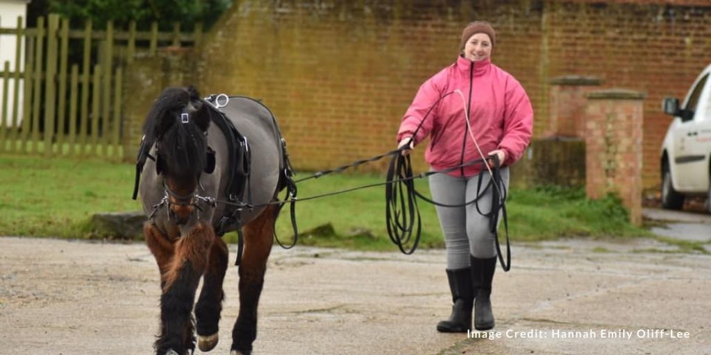
Over the last few years, I have found myself more and more strongly believing that horses should not be in work before they achieve skeletal maturity. Like you, I pride myself on being evidence based in my approach to life. And so when I set out to write this article, I had a pretty good idea what I was going to write. Turns out I was wrong.
Numerous risk factors can lead to catastrophic injury in the racehorse, including the age at which the animal starts training and the kind of exercise they are performing. Horses that start training at an older age, those that are trained for long distances at high speed, and those that are given intra-articular injections are all at a higher risk for catastrophic injury.
An article published in Animals, ‘Training Young Horses: The Science Behind the Benefits’, shares the evidence that supports starting horses in training from a young age. A Research Refresh is available in the Onlinepethealth Equine Members library.
Let’s dive into the evidence from that paper, together with the limitations of research in this particular area, and a few thoughts!
Breaking the Apollo curse
The Apollo curse gets its name from Apollo, the last winner of the Kentucky Derby to have won the race without having started his racing career as a two-year old. It took 136 years before Justify broke the curse in 2018, winning not only the Kentucky Derby, but claiming the full Triple Crown after entering his first race only a few weeks earlier.
Justify’s win sparked and re-opened the debate about the age at which racehorses should start their career, and of course, here we are having this discussion again.
What do we learn from the so-called Apollo curse? Over the 136 years between Apollo’s win and Justify’s win, many believed that a horse unraced as a two-year old could not win, while other top trainers dismissed the belief and raced their late-starters regardless. Over 60 horses have raced in the Kentucky Derby as unraced two-year olds; eight placed second or third, but none of them won.
A quick search on where Justify went after his winning streak reveals that he was retired undefeated several weeks later. In September 2019, ‘it was revealed via The New York Times that Justify tested positive for scopolamine, a banned substance that can be performance enhancing, in excess of what is normally found in tainted feed. The positive came just a few days after the Santa Anita Derby and would have normally resulted in a disqualification but the California Horse Racing Board ultimately dismissed the case’ (Wikipedia).
The benefits of waiting
The story about Justify is certainly interesting, and may have added fuel to the debate, but at the end of the day what we really want to know is whether or not starting training at an early age has a detrimental effect on soundness, longevity and performance.
The primary argument made for waiting to back and train young horses is based on the closure rates of the epiphyseal plates, with the spinal epiphyseal plates closing after 5 ½ years of age or later. Spinal integrity is essential for soundness, neurological integrity, and locomotion of the horse.
Anecdotally, many of us also feel there is a link between starting horses in competitive training from a young age, and conditions such as hock arthritis or proximal suspensory disease occurring later in life.
The article in Animals cited above examined the available literature on the topic, which makes it a more authoritative source than anecdotal findings, or our beliefs. The authors looked first at epidemiological studies in the racing industry, and then evaluated our understanding of musculoskeletal tissues and their response to training and mechanical stress.
Epidemiological studies
In 2003, Stover released a review of research in the racing industry confirming that musculoskeletal injuries are indeed the greatest cause of racehorse turnover in the industry. They also concluded that two-year old racehorses were not at an increased risk of injury compared to horses older than two, and that horses older than four were at a greater risk of injury than younger horses.
Exercise was shown to be a risk factor for injury occurrence, with horses that performed a single long-distance, high-speed run as exercise being at greater risk than horses that performed short-distance, high-speed runs. Consistent work also reduced the risk of injury; horses that exercised daily had fewer injuries than horses that had periods of rest as a result of lay-ups.
A study looking at racing thoroughbreds and standardbreds showed that horses that started race training at two had more race starts and greater earnings than horses that started race training later in life, regardless of whether the horse started to compete at two or not – it was the start of training that had a positive impact (Rogers et al., 2012).
Another study showed that thoroughbreds that started racing at two had more lifetime wins, money earned and years racing than horses that started later in life (Santschi et al., 2017).
An Australian study showed that thoroughbreds that started racing at a later age were at increased risk of retiring from racing. The more race starts a horse had as a two-year old, the lower the risk of retirement and the greater the average distance raced. This paper showed an inverse relationship between the age that the racing career starts, and the length of the career (Velie et al., 2013).
A meta-analysis of the risk factors for catastrophic musculoskeletal injury showed that exercise and age were two horse-level risk factors, and that the older the horse was when starting, the greater the risk of catastrophic injury (Hitchens et al., 2019).
An evaluation of British race-day fractures showed that the risk of fracture increases with age, and that the risk of fracture is higher in the first year of racing (Rosanowski, 2019).
Now let’s shift gears a little, and look at dorsal metacarpal disease, or bucked shins. In total, 70% of racehorses are affected in early training, and two-year old’s seem to be at greater risk than older horses (Nunamaker, 1990). Dorsal metacarpal disease is characterised by stress fractures in the dorsal cortex of the third metacarpal, partially caused by a lag time between bone resorption and bone formation during remodelling. This common occurrence is thought to be linked to the lifestyle change that comes with race training, including being removed from pasture, being stabled, and not being afforded voluntary exercise at speed (Hoekstra, 1990; Ross, 2010).
What about the epiphyseal plates? A study focused on the distal radial epiphyseal plates looked at a group of two-year olds with open, intermediate and closed epiphyseal plates. At the end of the racing season, 77% of the horses that started with open epiphyseal plates remained sound, while only 55-56% of horses with intermediate or closed plates remained sound.
The authors stated, ‘Many horses with open epiphyses raced six or more times and remained sound while numerous horses with closed epiphyses became unsound before their first race or before completing six races.
Their conclusion was that horses with closed epiphyseal plates showed a greater incidence of lameness and poor performance than those who started training with open epiphyseal plates.
Is the data we have, reliable?
The data above starts to draw a picture, but before we go any further, let’s consider some of the limitations of these studies. Firstly, they are observational. While they may demonstrate associations, they cannot prove causation. Many factors differ between horses that start training early and those that start later, including conformation, temperament, nutrition, environment, management, trainer philosophy and owner expectations. This creates a selection bias that may influence the results from the outset. For example, horses that are naturally more athletic and remain sound during their early development are more likely to enter training at a younger age, while horses with conformational abnormalities, slower rates of maturation or early injuries are more likely to have their training delayed.
Secondly, the study populations are largely limited to Thoroughbred racehorses and Standardbreds. These are highly selected populations, and we cannot automatically extrapolate the findings to the broader equine population or to horses competing in other disciplines.
Finally, while some studies evaluate longevity within a racing career, very few follow horses beyond retirement from racing. As a result, they provide little information on long-term musculoskeletal health, soundness or performance later in life. A horse may enjoy a long and successful racing career while developing pathology that only becomes clinically relevant years after retirement.
Bone
There are two processes occurring in bone: bone modelling and bone remodelling. Bone modelling occurs during the growth and development of the horse, while remodelling is a response to injury or a renewal of old bone. Bone modelling is responsible for helping the bone adapt to stresses and forces, and is most active in the first few years of life. While still present in the mature horse, it occurs more slowly in older horses. For this reason, the skeletal structure of a young horse is highly influenced by their environment and the daily stresses they experience. Short-term dynamic exercise as an adolescent has been shown to result in beneficial changes in bone morphology, increased fracture force, and reduced fracture risk at maturity (Hitney, 2004; Hitney, 2004; Warden, 2007; Logan, 2019).
Articular cartilage
Articular cartilage provides a smooth, lubricated surface for articulation, reduced friction and load transmission. Damage or trauma will lead to the development of OA, as there is a reduced healing and regenerative capability in this tissue.
Cartilage health is dependent on regular loading and movement, but strenuous long-term activity can lead to undesirable cartilage changes. Modifications to cartilage occur in response to high strain and vigorous exercise, and are site specific (Murray 2001). Both immobilisation and excessive exercise have been shown to be detrimental to the cartilage in young foals (Van De Lest, 2002).
We need more research to determine how much exercise is beneficial and how much is detrimental – and at what age.
Tendons and ligaments
The superficial digital flexor tendon (SDFT) and suspensory ligament (SL) are the main energy storing structures in the forelimb, and work close to their breaking point during high speed gallops. Nearly 30% of racehorses suffer an injury to the SDFT (Rich, 2014), and tendon healing is less than optimal, with the healed tendon retaining a reduced elasticity and reduced tensile strength.
At a young age, tendons have the capability to adapt to force, but lose that capacity as they age (Dowling, 2005; Docking, 2012). At 19 months of age, high-intensity treadmill exercise leads to an increased cross sectional area (CSA) of the deep digital flexor tendon (DDFT), but no change in the SDFT, suggesting that by this age, the SDFT has lost its adaptability (Dowling, 2005).
The risk of injury to the SDFT also increases with age (Patterson-Kane, 2012).
In foals, the SDFT doubles in size from the age of 50 days to one year, and reaches maturity at the age of two, while the DDFT and common digital extensor (CDE) undergo hypertrophy between two and three years of age (Birch 1999; Moffat, 2008). This raises the question of whether the SDFT can be strengthened through exercise, at a younger age. Foals with free access to pasture and spontaneous exercise were compared to foals with free access to pasture and forced exercise. There was no difference in the CSA of the SDFT between the two groups either at 12 months, or at two years when they started race training (Moffat, 2008).
Both a lack of exercise and excess exercise in young horses can negatively impact the tendon structure and function at maturity (Patteron-Kane, 2012; Smith 1999; Cherdchutham, 2010).
Age is not the only factor
‘While the age of horses entering training has been blamed for catastrophic injuries, the real culprit may be how we manage horses that are developing injuries’ (Logan, 2021).
Monitoring training, giving adequate rest and understanding the adaptability and adaptation of musculoskeletal structures may be key to preventing injury in the racehorse. Thermography may provide a valuable tool to allow us to detect problems before injury occurs in the young racehorse, and help us to respond appropriately.
Horses with minor injuries are more likely to be medically treated, so that the horse remains in training, than to be rested. Thoroughbreds and quarter horses that died as a result of race training had a five-times greater chance of having had hyaluronic acid injections than animals that did not (Hitchens, 2018). Horses that received corticosteroids up to 398 days before a race were at a greater risk of injury than untreated horses, and subsequent injections increased that risk. Introducing a cut-off of 30 days before a race, and ten days before training for any intra-articular corticosteroid injections, resulted in a dramatic decrease in catastrophic injuries (Paulick Report).
Conclusion
While we don’t yet have all the answers, particularly regarding long-term musculoskeletal health beyond a racing career, the evidence currently available challenges the long-held assumption that delaying training is always the safer option. Epidemiological studies consistently associate an earlier introduction to race training with reduced injury rates, longer racing careers and improved performance, while research on bone, cartilage, tendons and ligaments demonstrates that developing tissues respond positively to appropriate mechanical loading.
The evidence suggests that a gradual, carefully managed introduction to the mechanical stresses horses will encounter later in life allows bones, tendons, ligaments and cartilage to adapt in ways that become increasingly limited with maturity. Rather than asking simply when a young horse should begin work, perhaps we should instead be asking what that work should look like. How much exercise is enough to stimulate healthy adaptation? How much becomes excessive? And how can we progressively prepare the horse for the demands of its future career? What if, by waiting to introduce appropriate work, we are in fact increasing the risk of injury later in life?
Whatever age a horse begins training, the available evidence suggests that progression, monitoring and management are just as important as timing. Appropriate loading must be balanced with adequate recovery to allow musculoskeletal tissues to adapt while minimising the risk of overload. A veterinary team, including a qualified Vetrehabber, can play an important role in monitoring the horse throughout this process, identifying early signs of maladaptation and helping to guide training decisions that prioritise both performance and long-term welfare.
References
- CHRB approves continuing education program for trainers; poised to further tighten corticosteroid, thyroxin use—Horse Racing News | Paulick Report. Available online: https://www.paulickreport.com/news/the-biz/chrb-approves-continuingeducation-program-for-trainers-poised-to-further-tighten-corticosteroid-use/
- Hitchens, P.L., Hill, A.E. & Stover, S.M. (2018). Relationship between historical lameness, medication usage, surgery, and exercise with catastrophic musculoskeletal injury in racehorses. Vet. Sci. 5, 217.
- Hitchens, P.L., Morrice-West, A.V., Stevenson, M.A. & Whitton, R.C. (2019). Meta-analysis of risk factors for racehorse catastrophic musculoskeletal injury in flat racing. J. 245, 29–40.
- Hoekstra, K.E., Nielsen, B.D., Orth, M.W., Rosenstein, D.S., Schott, H.C. & Shelle, J.E. (1999). Comparison of bone mineral content and biochemical markers of bone metabolism in stall- vs. pasture-reared horses. Equine Vet. J. Suppl. 30, 601–604.
- Logan, A.A. & Nielsen, B.D. (2021). Training Young Horses: The Science behind the benefits. Animals, 11, 463. https://doi.org/10.3390/ ani11020463
- Nunamaker, D.M., Butterweck, D.M. & Provost, M.T. (1990). Fatigue fractures in thoroughbred racehorses: relationships with age, peak bone strain, and training. J. Orthop. Res. 8, 604–611.
- Rogers, C.W., Bolwell, C.F., Tanner, J.C. & Van Weeren, P.R. (2012). Early exercise in the horse. Vet. Behav. 7, 375–379.
- Rosanowski, S.M., Chang, Y.M., Stirk, A.J. & Verheyen, K.L.P. (2019). Epidemiology of race-day distal limb fracture in flat racing thoroughbreds in great britain (2000–2013). Equine Vet. J. 51, 83–89.
- Ross, M. & Dysonn, S. (2010). Diagnosis and management of lameness in the horse. 2nd ed. Amsterdam, The Netherlands: Elsevier.
- Santschi, E.M., White, B.J., Peterson, E.S., Gotchey, M.H., Morgan, J.M. & Leibsle, S.R. (2017). Forelimb conformation, sales results, and lifetime racing performance of 2-year-old thoroughbred racing prospects sold at auction. Equine Vet. Sci. 53, 74–80.
- Stover, S.M. (2003). The epidemiology of thoroughbred racehorse injuries. Tech. Equine Pract. 2, 312–322.
- Velie, B.D., Knight, P.K., Thomson, P.C., Wade, C.M. & Hamilton, N.A. (2013). The association of age at first start with career length in the australian thoroughbred racehorse population. Equine Vet. J. 45, 410–413.
- https://en.wikipedia.org/wiki/Justify_(horse)
Bone references
- Hiney, K.M., Nielsen, B.D. & Rosenstein, D. (2004). Short-duration exercise and confinement alters bone mineral content and shape in weanling horses. Anim. Sci. 82, 2313–2320.
- Hiney, K.M., Nielsen, B.D., Rosenstein, D., Orth, M.W. & Marks, B.P. (2004). High-intensity exercise of short duration alters bovine bone density and shape. Anim. Sci. 82, 1612–1620.
- Logan, A., Nielsen, B., Robison, C., Manfredi, J., Schott, H., Buskirk, D. & Hiney, K. (2019). Calves, as a model for juvenile horses, need only one sprint per week to experience increased bone strength. Anim. Sci. 97, 3300–3312.
- Nielsen, B.D., Potter, G.D., Morris, E.L., Odom, T.W., Senor, D.M., Reynolds, J.A., Smith, W.B. & Martin, M.T. (1997). Changes in the third metacarpal bone and frequency of bone injuries in young quarter horses during race training—observations and theoretical considerations. Equine Vet. Sci. 17, 541–549.
- Warden, S.J., Fuchs, R.K., Castillo, A.B., Nelson, I.R. & Turner, C.H. (2007). Exercise when young provides lifelong benefits to bone structure and strength. Bone Miner. Res. 22, 251–259.
Articular cartilage references
- Brama, P.A.J., Tekoppelle, J.M., Bank, R.A., Barneveld, A., Firth, E.C. & Van Weeren, P.R. (2000). The influence of strenuous exercise on collagen characteristics of articular cartilage in thoroughbreds age 2 years. Equine Vet. J. 32, 551–554.
- Murray, R.C., Birch, H.L., Lakhani, K. & Goodship, A.E. Biochemical composition of equine carpal articular cartilage is influenced by short-term exercise in a site-specific manner. (2001). Cartil. 9, 625–632.
- Van de Lest, C., Brama, P.A.J., Van Weeren, P., Van de Lest, C.H.A., Brama, P.A.J.& Ren Van Weeren, P. (2002). The Influence of exercise on the composition of developing equine joints. 39, 183–191.
- Van Weeren, P.R., Firth, E.C., Brommer, H., Hyttinen, M.M., Helminen, H.J., Rogers, C.W., DeGroot, J. & Brama, P.A.J. (2008). Early exercise advances the maturation of glycosaminoglycans and collagen in the extracellular matrix of articular cartilage in the horse. Equine Vet. J. 40, 128–135.
Ligaments/tendons
- Birch, H.L., McLaughlin, L., Smith, R.K. & Goodship, A.E. (1999). treadmill exercise-induced tendon hypertrophy: assessment of tendons with different mechanical functions. Equine Vet. J. Supp. 30, 222–226.
- Buchanan, C.I. & Marsh, R.L. (2002). Effects of exercise on the biomechanical, biochemical and structural properties of tendons. Biochem. Phys. 133, 1101–1107.
- Cherdchutham, W., Becker, C., Smith, R.K.W., Barneveld, A. & Weeren, P.R. (2010). Age-related changes and effect of exercise on the molecular composition of immature equine superficial digital flexor tendons. Equine Vet. J. 31, 86–94.
- Docking, S.I., Daffy, J., Van Schie, H.T.M. & Cook, J.L. (2012). Tendon structure changes after maximal exercise in the thoroughbred horse: use of ultrasound tissue characterisation to detect in vivo tendon response. J. 194, 338–342.
- Dowling, B.A. & Dart, A.J. (2005). Mechanical and functional properties of the equine superficial digital flexor tendon. J. 170, 184–192.
- Moffat, P.A., Firth, E.C., Rogers, C.W., Smith, R.K.W., Barneveld, A., Goodship, A.E., Kawcak, C.E., McIlwraith, C.W. & Weeren, P.R. (2008). The influence of exercise during growth on ultrasonographic parameters of the superficial digital flexor tendon of young thoroughbred horses. Equine Vet. J. 40, 136–140.
- Patterson-Kane, J.C., Becker, D.L. & Rich, T. (2012). The pathogenesis of tendon microdamage in athletes: the horse as a natural model for basic cellular research. Comp. Pathol. 147, 227–247.
- Rich, T. & Patterson-Kane, J.C. (2014). Science-in-brief: What is needed to prevent tendon injury in equine athletes? A conversation between researchers and industry stakeholders. Equine Vet. J. 46, 393–398.
- Smith, R.K., Birch, H., Patterson-Kane, J., Firth, E.C., Williams, L., Cherdchutham, W., Van Weeren, W.R. & Goodship, A.E. (1999). Should equine athletes commence training during skeletal development? changes in tendon matrix associated with development, ageing, function and exercise. Equine Vet. J. 30, 201–209.
0 Comments