

Equine headshaking can be an extremely challenging condition to treat and manage, and in many cases causes significant distress to both the horse and owner. It can make horses dangerous and unrideable, and may be the reason a horse is put to sleep.
As our understanding of trigeminal-mediated headshaking has improved, we have developed more targeted approaches to diagnosis and management. Despite these advances, the condition remains one of the most frustrating disorders encountered in equine practice.
Let’s explore equine headshaking in light of new research published in this area.
What is Headshaking?
Trigeminal-mediated headshaking (TMHS) is now recognised as the most common form of equine headshaking. It is characterised by trigeminal nerve sensitisation, resulting in neuropathic facial pain that may feel similar to burning, tingling, itching or electric shock-like sensations described by human patients).
Headshaking presents in horses most commonly as a drop and sharp flick or jerk of the head in the vertical plane that can be quite violent. It can also include flicking or jerking in the horizontal plane, rubbing of the muzzle, striking towards their face with their forelimbs, snorting, and is often coupled with an anxious expression (Aleman & Morales, 2026).
Clinical signs vary considerably in frequency, severity and duration between horses. They may remain year round, or occur seasonally in the spring and summer.
Some of the triggers of headshaking can be bright sunlight (photic headshaking), wind stimulating the muzzle, eating dry foliage, exercise, sharp loud noise, pollen and seasonal allergens, and flies around the face.
Trigeminal Nerve Anatomy
The trigeminal nerve is the largest cranial nerve and provides sensory innervation to much of the face through three major divisions: the ophthalmic (V1), maxillary (V2) and mandibular (V3) nerves. The mandibular nerve also carries motor fibres to the muscles of mastication.
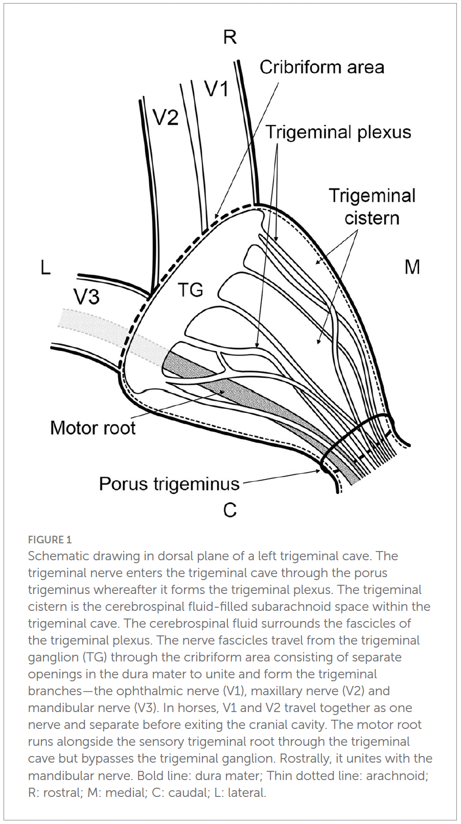
The trigeminal nerve originates from the brainstem and enters the trigeminal cave, a dural recess within the cranial cavity. Within the trigeminal cave, the nerve forms the trigeminal plexus before converging rostrally into the trigeminal ganglion. Becker et al. (2024) demonstrated that, in horses, the trigeminal ganglion and nerve fascicles are surrounded by a cerebrospinal fluid-filled subarachnoid space, forming the trigeminal cistern. From the trigeminal ganglion, nerve fascicles pass through the dura and unite to form the three major divisions of the trigeminal nerve – the ophthalmic (V1), maxillary (V2) and mandibular (V3) nerves (Becker et al. 2024).
In the horse, the ophthalmic nerve (V1) passes towards the orbit through the orbital fissure. The maxillary nerve (V2) leaves the cranial cavity through the round foramen (foramen rotundum), before continuing towards the face and ultimately giving rise to branches supplying structures including the nose, nostrils, upper lip, upper teeth and maxillary sinuses. The mandibular nerve (V3) leaves the skull through the oval notch of the foramen lacerum and provides both sensory and motor innervation (Budras et al., 2009).
There is another important neurological relationship to consider: the convergence of cervical and trigeminal sensory afferents.
Within the central nervous system, sensory information from the trigeminal nerve does not exist independently from sensory information arising from the upper cervical region. Trigeminal nociceptive afferents and afferents from the upper cervical spinal nerves converge onto second-order neurons within the trigeminocervical complex. This convergence provides a mechanism through which nociceptive input from structures innervated by C1–C3 may influence trigeminal sensory processing, and vice versa (Bogduk, 1992; Piovesan et al., 2003).
Trigeminal Nerve Involvement
There has been confirmation of the involvement of the trigeminal nerve through sensory nerve conduction tests evoking action potentials of the trigeminal nerve. In two studies (Aleman et al., 2013, 2014), 6 control horses were compared to 6 headshakers through sensory nerve conduction tests.
They found that the headshaking horses had a lower stimulus threshold than the control group, with bilateral nerves being equally affected. There were no other differences in neurophysiological parameters, such as conduction velocity.
One headshaker in the study was seasonally affected and was in remission at the time of testing. For this horse, the stimulus threshold was identical to normal horses, suggesting that the nerve stimulus threshold is malleable and can be returned to normal ranges.
Together, these findings support the current understanding that TMHS is a functional disorder characterised by trigeminal nerve sensitisation, rather than a disease caused by structural nerve damage.
Why Does the Trigeminal Nerve Become Sensitised?
While research has demonstrated altered trigeminal nerve excitability in horses with TMHS, the question of why sensitisation occurs remains unanswered.
Dr Audrey DeClue draws attention to the relationship between headshaking and exercise, and questions whether the typical age of presentation may coincide with increasing ridden workload, training and competition. In her clinical experience, she describes marked poll sensitivity, upper cervical pain and positional headshaking, where changing the position of the head and neck can trigger or alter clinical signs.
DeClue’s treatment approach focuses on relieving pain within the upper cervical region, particularly C1–C3, and she reports a high level of clinical success with this approach. While this does not establish upper cervical pain as the cause of TMHS, it raises an interesting question when considered alongside the convergence of cervical and trigeminal sensory afferents. Could reducing nociceptive input from C1–C3 influence trigeminal sensory processing through the trigeminocervical complex?
DeClue’s clinical observations provide an interesting reason to consider the upper cervical region – and the relationship between cervical pain and trigeminal signalling – more closely in horses presenting with headshaking.
Listen to The Horse First Podcast on Headshaking Syndrome
Diagnosis of Trigeminal-Mediated Headshaking (TMHS)
TMHS is a diagnosis of exclusion, meaning there is no single test that can definitively confirm the condition (Pickles, 2023). Instead, diagnosis relies on recognising the characteristic clinical signs, obtaining a thorough history, and systematically ruling out other causes of headshaking behaviour. A complete physical examination should be supported by detailed oral, ophthalmic and otoscopic examinations, with additional diagnostic imaging or endoscopy performed where indicated to investigate dental disease, sinus pathology, upper airway disorders, ear disease or other sources of facial pain. Musculoskeletal dysfunction, cervical pain and poor tack fit may produce similar clinical signs or coexist with TMHS, and should therefore be assessed during the diagnostic work-up. Once alternative causes of headshaking have been excluded, TMHS may be considered as the likely diagnosis (Aleman & Morales, 2026).
Treatment and Management of TMHS
As the cause of TMHS remains unknown, treatments aim to reduce sensitization of the trigeminal nerve. These include dietary modifications and physical protection. Additionally medical and surgical interventions have been explored and implemented. Management strategies should always be implemented based on the needs of the individual horse (Aleman & Morales, 2026).
Physical Protection: Nose Nets and UV Protective Masks
When headshaking is triggered by photosensitivity or light, a facemask with UVA and UVB protection that blocks at least 90% of UV rays can be helpful to reduce light-induced stimulation of the trigeminal nerve. Both goggles and masks are available, and 50% of owners have reported a successful outcome from their use (Pickles et al., 2014). Photo-sensitivity may not be the only trigger present in horses.
Nose nets can be a simple, effective management strategy. In theory, nose nets alter the sensory stimulation to through gentle contact with the nose. With various kinds of nose nets available, it is recommended to try multiple styles and designs before moving on to a new management strategy (Aleman & Morales, 2026).
Dietary Modifications
Modifications to the diet can be used to support other management practices, but are rarely effective on their own. Increasing the pH of the diet should be the first change. This can be achieved by reducing grain concentrate while adding beet pulp and lucerne to the diet. Theoretically, this change in pH may increase the firing threshold of the trigeminal nerve.
The microbiota of the gut may play a role, and is an area of emerging interest.
Magnesium has been investigated as an adjunctive treatment because of its effects on neuronal excitability. It interacts with GABA receptors in an inhibitory role, and blocks NMDA receptors to prevent glutamate release. Providing boron alongside magnesium can improve the availability of magnesium (Sheldon et al., 2019).
Electrical Therapy
Electrotherapy can be used to alter nerve conduction and reduce pain, through transcutaneous or implanted electrical devices. The most commonly used methods include TENS, PENS, electroacupuncture or transmagnetic stimulation.
We are familiar with transcutaneous electrical nerve stimulation (TENS), but Percutaneous electrical nerve stimulation (PENS) may be a new term to you.
With the application of PENS, a conductive needle is inserted into the tissue to directly stimulate the nerve. It is recognized as a treatment for human patients with nerve pain, and provides relief to approximately 70% of patients with postherpetic or post-traumatic trigeminal neuropathic pain (Johnson & Burchiel 2004).
In equines, PENS has shown a 50% success rate (Roberts et al., 2016, 2017) with reduced symptoms (or complete remission) for an average of 15 weeks, at which point the treatment can be repeated. Although PENS currently has the strongest evidence among neuromodulation techniques, response rates remain variable and repeat treatments are often required.
Devereaux 2017 has reported some success with the use of electroacupuncture, which may be more readily available than PENS.
Conclusion
Trigeminal-mediated headshaking remains one of the most challenging neurological conditions encountered in equine practice. Although the underlying cause of trigeminal nerve sensitisation is still unknown, our understanding of the disorder has advanced considerably over the past two decades. We now recognise TMHS as a functional neuropathic pain disorder, allowing clinicians to focus on identifying characteristic clinical signs, ruling out other causes of headshaking, and implementing an individualised management plan based on each horse’s specific triggers. While no single treatment is universally effective, combining environmental management, physical protection, dietary modification and selected medical or neuromodulation therapies can improve comfort and quality of life for many horses.
References
Aleman, M. and Morales, C.J. (2026) ‘Update on idiopathic trigeminal-mediated headshaking’, Veterinary Clinics of North America: Equine Practice, 42(1), pp. 117–131. https://doi.org/10.1016/j.cveq.2025.12.003
Aleman, M., Pickles, K.J., Simonek, G. and Madigan, J.E. (2012) ‘Equine herpesvirus-1 in trigeminal ganglia of horses with idiopathic headshaking’, Journal of Veterinary Internal Medicine, 26, pp. 192–194.
Aleman, M., Williams, D.C., Brosnan, R.J., Nieto, J.E., Pickles, K.J. et al. (2013) ‘Sensory nerve conduction and somatosensory evoked potentials of the trigeminal nerve in horses with idiopathic headshaking’, Journal of Veterinary Internal Medicine, 27, pp. 1571–1580.
Becker, R., Haenssgen, K., Precht, C., Khoma, O.Z., Hlushchuk, R., Koch, C., Kaessmeyer, S. and de Preux, M. (2024) ‘An anatomical study of the subarachnoid space surrounding the trigeminal ganglion in horses—in preparation for a controlled glycerol rhizotomy in equids’, Frontiers in Veterinary Science, 11, 1424890. https://doi.org/10.3389/fvets.2024.1424890
Bogduk, N. (1992) ‘The anatomical basis for cervicogenic headache’, Journal of Manipulative and Physiological Therapeutics, 15(1), pp. 67–70.
Budras, K.D., Sack, W.O. and Röck, S. (2009) Anatomy of the Horse: An Illustrated Text. 5th edn. Hannover: Schlütersche.
Devereux, S. (2017) ‘Electroacupuncture as an additional treatment for headshaking in six horses’, Equine Veterinary Education.
Johnson, M.I. and Burchiel, K.J. (2004) ‘Peripheral stimulation for treatment of trigeminal postherpetic neuralgia and trigeminal posttraumatic neuropathic pain: A pilot study’, Neurosurgery, 55(1), pp. 135–141. https://doi.org/10.1227/01.NEU.0000126874.08468.89
Madigan, J.E. and Bell, S.A. (2001) ‘Owner survey of headshaking in horses’, Journal of the American Veterinary Medical Association, 219, pp. 334–337.
Pickles, K. (2019) ‘Is electrical nerve stimulation the answer for management of equine headshaking?’, Veterinary Clinics of North America: Equine Practice, 35(2). https://doi.org/10.1016/j.cveq.2019.03.002
Pickles, K. (2023) ‘Trigeminal-mediated headshaking: A diagnostic challenge’, Equine Veterinary Education, 35, pp. 195–196. https://doi.org/10.1111/eve.13723
Pickles, K.J., Berger, J., Davies, R., Roser, J. and Madigan, J.E. (2011) ‘Use of gonadotrophin releasing hormone immunisation in equine headshaking’, Veterinary Record, 168(1), pp. 19–22.
Pickles, K.J., Madigan, J.E. and Aleman, M.R. (2014a) ‘Idiopathic headshaking: Is it still idiopathic?’, The Veterinary Journal, 201, pp. 21–30.
Pickles, K.J., Madigan, J.E. and Aleman, M.R. (2014b) ‘Owner-reported response to treatment in 130 headshaking horses’, Proceedings of the American Association of Equine Practitioners.
Pickles, K.J., Marlin, D.J., Williams, J.M. and Roberts, V.L.H. (2025) ‘Use of a poll-mounted accelerometer for quantification and characterisation of equine trigeminal-mediated headshaking’, Equine Veterinary Journal, 57(3), pp. 645–653. https://doi.org/10.1111/evj.14132
Piovesan, E.J., Kowacs, P.A. and Oshinsky, M.L. (2003) ‘Convergence of cervical and trigeminal sensory afferents’, Current Pain and Headache Reports, 7(5), pp. 377–383. https://doi.org/10.1007/s11916-003-0037-x
Roberts, V.L., Fews, D., McNamara, J.M. and Love, S. (2017) ‘Trigeminal nerve root demyelination not seen in six horses diagnosed with trigeminal-mediated headshaking’, Frontiers in Veterinary Science, 4, Article 72.
Roberts, V.L.H., Patel, N.K. and Tremaine, W.H. (2016) ‘Neuromodulation using percutaneous electrical nerve stimulation for the management of trigeminal-mediated headshaking: A safe procedure resulting in medium-term remission in five of seven horses’, Equine Veterinary Journal, 48(2), pp. 201–204.
Sheldon, S.A., Aleman, M., Costa, L.R.R. et al. (2019) ‘Effects of magnesium with or without boron on headshaking behaviour in horses with trigeminal-mediated headshaking’, Journal of Veterinary Internal Medicine, 33, pp. 1464–1472.









0 Comments