
A quick search in the Small Animal Vetrehabbers Facebook community shows eight posts where professionals have asked for advice about Tethered Cord Syndrome patients, or where TCS has come up in the case discussions where Vetrehabbers have become stuck with a particular clinical case.
A quick search on google scholar reveals that more than seven papers have been published in the last six years on TCS in canines.
While it may feel like this condition has sprung up out of the blue – and now every 5th patient is suspected of having it – this isn’t a new condition, and there is a good chance that it is even more prevalent in the canine population than we are yet recognizing.
This blog will provide a quick reference point for veterinary rehabilitation professionals who are unfamiliar with this condition. We discuss what it is, how it might present, the diagnostic procedure, surgical correction, and finally the experience of the Veterinary Rehabilitation Community in the treatment of this condition in their clinical practices.
The first part of this blog focuses on the most recent retrospective case series published by Lampe et al., 2026. They were able to include 12 dogs with a confirmed diagnosis and surgical intervention in the report.
The second part of this blog shares the experiences of Veterinary Rehabilitation Therapists globally, who have had been working with these cases.
What is Tethered Cord Syndrome?
TCS, or Tethered Cord Syndrome, occurs when the end of the spinal cord becomes tethered or adhered in the lumbar spine. This results in a lack of movement of the spinal cord within the vertebral canal as the spine flexes and extends during normal activity.
In humans, TCS can occur as a result of a mechanical adhesion, or when there is a loss of elasticity in the normal structures connecting to the spinal cord (Abdulrazeq et al., 2024).
In canines, TCS may occur on it’s own or in conjunction with congenital defects of the spine or spinal cord (Roynard & Dewey, 2025).
A Recap of the Anatomy Involved
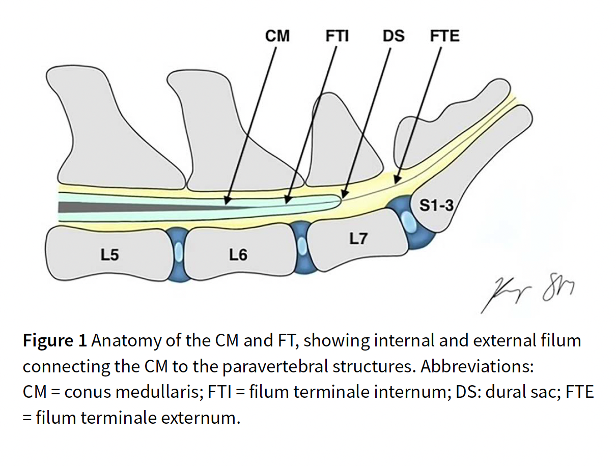
Understanding the anatomy that is affected is always the first step to understanding a condition like TCS and how it is treated.
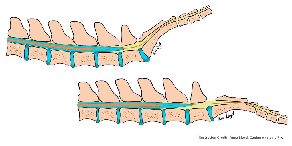
During growth, the spinal cord and the spinal column do not lengthen to the same degree. By the time a puppy reaches adulthood, the spinal cord within the spinal column ends around L6, while the nerve roots continue caudally to the level where they exit the spinal canal.
This is the reason that dorsal and ventral nerve roots along the length of the spine diverge from the spinal cord cranially from the point where they exit the spinal canal – we can see this cranial shift already occurring from the cervical region.
The spinal cord ends in the lumbar region as a tapered structure called the conus medullaris (latin for medullary cone). The exact level where the conus medullaris (CM) ends will vary depending on the size and breed of the dog.
The CM transitions into a thin strand of connective tissue called the filum terminale (latin for terminal thread) which continues caudally to attach onto a sacral or caudal vertebrae, acting as an anchor for the spinal cord (Fletcher, 2016).
The filum terminale has an internal and an external portion – determined by the dural sac. The dural sac surrounds the spinal cord and extends beyond the CM to create the lumbar cistern. The filum terminale starts internally, and continues externally to the dural sac.
Of course, the filum terminale is not the only structure that extends caudally in the vertebral canal – it is accompanied by the sacral and caudal nerve roots known collectively as the cauda equina.
In TCS, restrictions occur in this region that limit the glide of the CM within the spinal canal. This can occur if the filum terminale is thickened, shortened, inelastic, or attached abnormally to surrounding structures. It can also occur as a result of direct adhesions of the dural sac to the spinal canal (Lampe et al., 2026).
When adhesions occur, normal movements result in abnormal tension and traction on the spinal cord.
What Does TCS Look Like Clinically?
One of the most interesting findings from case reports and the community, is that dogs rarely present with any obvious pathology – no clear neurological deficits or orthopedic flags. Instead, many present as behavioural cases, pain cases, or with vague frustrating symptoms that don’t fit into any of our normal boxes.
Clinical signs often begin as subtle and gradually progress over months or years.
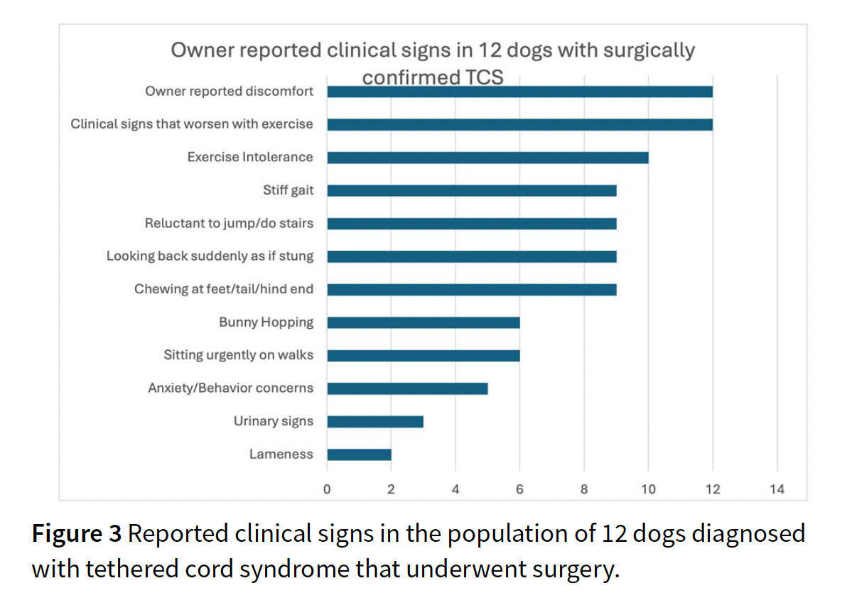
The most common owner reported signs point towards transient paresthesia, and include
- Suddenly looking at their hindquarters,
- Sitting urgently on a walk,
- Biting or chewing at their feet, tail or hindlimbs,
- Suddenly stopping during activity,
- Acting as if suddenly stung.
Additional owner reported signs that may be present include:
- Sensitivity to touch
- Vocalization
- Difficulty becoming comfortable
- Reduced exercise tolerance
- Reluctance to jump or use stairs
- Urinary dysfunction
On evaluation, Vetrehabbers may also identify:
- Stiff pelvic limb gait,
- Delayed hindlimb paw placement,
- Weak withdrawal reflexes,
- Pain on lumbar palpation, worsening when the vertebrae are palpated with hips elevated,
- Pain on hip extension,
Lampe et al., (2026) reported that dogs diagnosed with TCS had already seen specialists, rehabilitation therapists, and undergone pain control trials with no relief, before the diagnosis was reached.
“All the owners reported that their dogs’ clinical signs worsened with exercise, and reported an activity suggestive of transient paresthesia, such as looking suddenly at their hindquarters, chewing at the paw or tail, or sitting urgently while on walks” Lampe et al., (2026).
Diagnostic Testing for Tethered Cord Syndrome
TCS is particularly difficult to diagnose in dogs because the location of the conus medullaris varies greatly between dogs of different sizes and breeds. Additionally, static MRI or CT scans are likely to appear completely normal (Lampe et al., 2026).
For an accurate diagnosis, we need to measure whether the CM glides within the spinal canal in response to flexion and extension of the spine. To achieve this, dynamic MRI or CT scans are necessary (Lampe et al., 2026).
Surgical Intervention
Surgical treatment aims to release the structures that create abnormal tension on the spinal cord.
A lumbosacral dorsal hemilaminectomy allows identification and transection of any structures tethering the dural sac. The filum terminale is assessed for tension both internally and external and if needed a durotomy is performed to release it (Lampe et al., 2026).
The goal is to remove adhesion, reduce tension and allow the spinal cord to move normally again.
Post-op Care
Post-op recommendations may include a short period of hospitalization for pain management, followed by home analgesia, strict activity restriction, and gradually increasing controlled exercise over several weeks, depending on the surgeon’s protocol and the individual dog (Lampe et al., 2026).
Outcomes
According to case reports, post-op outcomes are positive with dogs showing improvements in their clinical signs. Transient paresthesia may completely resolve or significantly improve.
While some neurological deficit may occur in the short-term such as delayed paw placing, these deficits resolve or return to normal shortly after surgery. We can expect less back pain, improved posture and exercise tolerance, and a reduced need for analgesia. According to Lamp et al., (2026), all owners reported that their dogs had an improved quality of life.
How Vetrehabbers Have Intervened
David Lane, DVM, DACVSMR, CVSMT, CCRT, CVA
My experience with tethered cord syndrome (TCS) is that there are several primary clinical signs. Before I was aware of TCS, I had a number of cases I considered “lemons” – young, non-athletic dogs with recurrent lumbar pain. Once I became aware of TCS and saw a pattern, I referred these patients for imaging, and every single one was diagnosed with Tethered Cord Syndrome.
The classic case is a young dog, often less than 2 years old. The signs I look for include:
- Recurrent myofascial back pain, particularly lumbar pain, that is either refractory to normal treatment techniques, or recurs with unexpected frequency, with no identifiable provocation.
- nonspecific symptoms of back pain, such as sitting down on walks,
- poor exercise stamina,
- behavioral changes indicating defensiveness, anxiety, or aggression to protect the lower back,
- a skipping gait lameness,
- sudden looking or chewing at the back end.
It is important to emphasize that these signs generally start at a young age. Some dogs have mild, non-progressive symptoms and can be treated conservatively.
Pregabalin is an excellent pharmaceutical intervention for these dogs. If I am suspicious of TCS, I will perform a pregabalin therapeutic trial; a dramatic response substantially increases my suspicion of TCS.
For rehabilitation, I use my standard myofascial treatment techniques but with a gentler approach for TCS dogs. I find they are predisposed to rebound pain, maladaptive pain, and central sensitization, which explains the benefits of pregabalin.
Patients with a more severe clinical signs, or whose signs are worsening over time, are candidates for surgery. Post-operative rehabilitation is similar to that for a hemilaminectomy, with the key difference being the introduction of dural gliding. I understand dural gliding is a mainstay in treating TCS in humans, and we have had good success with it. We use a massage therapist who has had additional training in dural gliding; my understanding is that it requires a gentle touch.
A large percentage of surgical cases will have setbacks; the trajectory of post-operative healing is not a straight line. If a patient has a setback without significant myofascial pain, we have had the best luck with ketamine constant rate infusions (CRIs). I am not a proponent of subcutaneous ketamine to treat maladaptive pain, as in my experience, it has been ineffective. However, a round of 3 ketamine CRIs, using Lindsay Fry’s protocol, is quite effective. If you have a TCS case with a significant setback, I would be quick to go to a ketamine CRI. We are in the process of collecting data to hopefully have some numbers on its effectiveness in the future. There is a small amount of anecdotal evidence to suggest that naltrexone may have utility in some cases.
I have been in discussions with Dr. Rachel Lampe about doing pre-emptive ketamine CRIs to see if we can demonstrate a lower incidence of maladaptive pain setbacks. Because of this history of maladaptive pain, I am in no rush to take these patients off pregabalin post-operatively.
Theresa Miceli, BA BVMS MRCVS
Cases: 7
General presentation: varies. No really overt neurological symptoms but usually refractory back pain and sensitivity. Back pain can be localized at the LS junction but there can be compensatory back pain from weight shifting higher up as well. There are usually behavioural changes as well- increased reactivity with people or other dogs, touch sensitivity, light chasing, noise sensitivity, resource guarding.
Priorities in treatment: Central desensitization. All of these patients get low dose subcutaneous ketamine. The local neurologist also prefers Pregabalin over gabapentin. And I also find an NSAID helps. Possibly some methocarbamol as well. I haven’t come across one dog that hasn’t been helped so far with medication and treatment (my treatment also includes acupuncture, PEMF, NMES, tuning fork therapy, MFR, vibration therapy- and usually a referral to hydro).
Patterns: no orthopaedic reasons for pain as I usually try to rule these out first. There may also be one hind limb that has slightly different muscle mass compared to the other.
I think behavioural changes coupled with no orthopaedic reason for pain would be the two things that increase the suspicion of TCS and lead to alerting owners of the possibility.
Kirsty Powell-Palmer PgDip Vet Physio, BSc(hons) Vet Nurse, CCFT
I’ve tried to summarize an interesting case load. We are up to 10 cases in 3 months of diagnosed TCS, mainly because we have a neurologist who is happy to look into things beyond the obvious neurological conditions.
Over the past one to two years, I have seen a growing cohort of dogs that were initially referred for behavioural concerns – maladaptive pain responses, anxiety, reactivity, and poor progress with behavioural interventions.
Working collaboratively with a behavioural veterinarian, a behaviour modification trainer, and more recently a neurologist who has joined our province, we began to notice consistent physical patterns underlying these cases. Clinically, many of these dogs presented with subtle but repeatable findings: mild ataxia without clear conscious proprioceptive deficits, sciatic nerve pathway sensitivity, piriformis discomfort, reduced hind limb engagement, and lumbosacral pain. A significant number also had a history of early-onset behavioural signs, including intermittent anxiety, reactivity, and behaviours such as flank or groin-directed fly biting.
A major barrier to assessment was that these patients often poorly tolerated traditional veterinary examination. Many had never been fully examined while conscious. As a result, our primary focus became building trust through cooperative care. Sessions were intentionally slow, using non-traditional exam approaches – on the floor, at the dog’s pace, with ample reinforcement. Early assessment relied heavily on observation, dynamic movement analysis, and active range of motion, as palpation was often not initially possible. Muzzle use ensured safety while maintaining patient comfort. As trust developed over weeks to months, we were able to perform more complete examinations, consistently identifying reduced hind limb muscle mass, axial muscle loss, and persistent lower back pain.
Initial management often followed a conservative approach similar to low-grade IVDD: Class IV laser therapy, manual therapies (including massage and skin rolling), and targeted strengthening exercises such as weight shifting and sit mechanics. A key therapy throughout these cases has been Class IV laser. Its ability to target large areas quickly meant that even in patients who did not initially tolerate palpation, we could begin treatment as soon as a small amount of trust was established. The response has been consistently remarkable – many dogs visibly relaxing, softening, and in most cases falling asleep during treatment.
Despite the use of anti-inflammatories and neuropathic pain medications, the laser, with its direct and localized effect, has often provided the most significant and immediate relief.
One particularly notable case had never rested outside of his crate in his 18 months of life. He required enforced “time-outs” because he was unable to decompress independently. During his very first session, which was otherwise a challenging, high-arousal exam, we applied laser to his lumbar spine. He laid down, fell asleep, and did not move for 15 minutes. This was a major milestone for him – not only resting, but doing so in a novel environment. Moments like this have highlighted both the level of underlying discomfort these dogs may be experiencing and the impact of appropriate, targeted intervention.
Until recently, access to advanced imaging was limited, and cases without overt neurological deficits were not prioritized for MRI. With the recent availability of dynamic MRI, several of these patients have now been diagnosed with tethered cord syndrome (TCS), confirming a pattern we had been clinically suspecting. These cases are currently being further evaluated in collaboration with our neurologist and consulting specialists.
While some dogs are surgical candidates, others are being managed conservatively. For both groups, rehabilitation is expected to play a key role – supporting muscle recruitment, improving function, and assisting with pain management pre- and post-operatively.
For me, the biggest takeaway has been to slow down and really listen to the patient. These dogs didn’t come in as “neurological” cases – they were behavioural dogs who were scared, reactive, and often unable to be examined. By taking the time to build trust, adapting how we assess them, and working closely as a team, we were able to start seeing patterns that didn’t fit the usual boxes. It’s also made me realise how many of these cases may be out there if we’re willing to look a bit differently, especially in dogs showing subtle, pain-related behavioural changes.
Jenn Panko, RVT, CCRP, VTS Physical Rehabilitation, Cert OA Case Management, Cert Companion Animal Pain Management, FFCP
It can feel as though TCS is everywhere right now, which makes a thoughtful case-by-case approach even more important. I caution everyone to treat the dog in front of you. Pain management, including management and prevention of wind-up pain, is incredibly important. Don’t forget to treat the whole dog, including concurrent osteoarthritis, body condition, and unrelated musculoskeletal and neurological conditions.
Pain patterns can change after surgery as the nervous system and surrounding soft tissues adapt. I often describe it to owners as “retractable leash disease” — when the spinal cord suddenly has more freedom of movement, tissues that have adapted to longstanding tension may respond in unexpected ways. Concurrent shoulder, neck, pectoral, and jaw pain are common. Limb and tail chewing can come and go and should be monitored.
As a veterinary technician, it is important to work collaboratively with a clinician who can provide comprehensive pain management, including constant rate infusion therapy when indicated. Acupuncture, chiropractic care, and massage therapy have proven to be helpful in many cases. I frequently find pain and dysfunction between the scapulae and have found slow, progressive dural gliding exercises, combined with core strengthening and rehabilitation strategies that address kinetic chain dysfunction and muscle firing patterns, to be among the most effective interventions.
The real challenge is often aligning owner expectations and reducing fixation on day-to-day fluctuations. Recovery rarely follows a straight line and more often, progress looks like scribbles on a page that trend upward over time.
From the Professional Owners’ Perspective:
Sara Coeckelberghs, PT
Our little sausage dog Sky has had some issues from the very start. When she was 5 month old, she bumped into our other dachshund while playing, and lost consciousness. She was diagnosed with atlantoaxial instability and had surgery to fix it. Her rehab went okay, but something felt off. I kept worrying about her hind weakness and her anxious behavior. We went to see an ortho, who did x-Rays. Like all dachshunds, they found abnormalities in the joints, but nothing that could explain this pain, behavior and weakness. We want to a behavioral vet, she prescribed pain medicine, gabapentine and anxiety medicine. She seemed better, but never really ‘good’.
In September we took her on a long road trip to Norway and it was hell for her. Totally overwhelmed, barking at everything and nothing, not eating in 4 weeks. Coming back home, I went to see the vet in the clinic where they treated her neck. We had a double appointment with ortho and neuro. They did a scan and a dynamic scan, and found the tethered cord.
They told me, a surgery could be considered, but with no guaranties of re-attachment of the cord because of scar tissue. However they also saw an instability at LS level, so we are a bit anxious of her future.
For now, we are opting for conservative management: she’s on medication, stabilisation physio therapy with mommy, and we are limiting her activities unfortunately. We have to minimize physical stress as well as emotional stress.
I have to say, even as a physio, I find it really hard to cope with this condition!
Sarah Hedderly,
Sarah shares Sparkle’s story and raises multiple additional points that are not included in the beginning of this blog, where we discuss what we already ‘know’ from published literature.
Sparkle’s story highlights that there is more we don’t know:
- Occult Tethered Cord does not show on dynamic MRI,
- Sacral dimples could be an indication to look closer,
- Pay attention to UTI’s and gut dysfunction,
- Pay attention to changes in the coat in the pelvic and spinal areas.
For Sarah, her journey with Tethered Cord Syndrome unfortunately did not end with Sparkles. In 2026, her second dog, Beau, was diagnosed and treated.
While Beau had much more subtle symptoms, the following stood out to me from her story:
- Poor gut health from puppy,
- Developing meningitis arteritis at 12 weeks,
- Challenges with bowel and bladder,
- Abnormal posture in a down position,
- Sensitivity/reactivity, including noise sensitivity,
- Regurgitating water, and once vomiting blood.
When listed like this, these symptoms don’t seem subtle – but as Beau loves training and successfully competed in agility and obedience, the symptoms just didn’t make sense.
Learn more from Dr Rachel Lampe in our interview
References
Lampe R, Kalamkarova E, Barnard L, Mareschal A, Keenihan EK, Sharp NJH. Diagnosis, surgery, and outcome of tethered cord syndrome in 12 dogs, Journal of Veterinary Internal Medicine, Volume 40, Issue 1, January-February 2026, aalaf031, https://doi.org/10.1093/jvimsj/aalaf031
Abdulrazeq H, Leary OP, Tang OY, et al. The surgical histopathology of the filum terminale: findings from a large series of patients with tethered cord syndrome. ClinMed.2024; 13:6.https://doi.org/10.3390/jcm13010006
De Decker S, Gregori T, Kenny PJ, et al. Tethered cord syn-drome associated with a thickened filum Terminale in a dog. J VetInternMed.2015;29:405-409.https://doi.org/10.1111/ jvim.12522
Shamir M, Rochkind S, Johnston D. Surgical treatment of tethered spinal cord syndrome in a dog with myelomeningocele. Vet Rec.2001; 148:755-756.https://doi.org/10.1136/vr.148.24. 755
Roynard P, Dewey CW. Lumbosacral (myelo) meningoceles in dogs, related tethered cord syndrome, and their surgical management: review of the literature and clinical experience. FrontVetSci.2025; 12:1510800.https://doi.org/10.3389/fvets. 2025.1510800
Espinosa, Romero J, De Decker S, Santifort K, et al. Occult tethered cord syndrome: insights into clinical and MRI features, prognostic factors, and treatment outcomes in 30 dogs with confirmed or presumptive diagnosis. FrontVetSci.2025; 12.https://doi.org/10.3389/fvets.2025.1588538
De Decker S, Watts V, Neilson DM. Dynamic lumbosacral magnetic resonance imaging in a dog with tethered cord syndrome with a tight filum terminale. Front VetSci.2017; 4:27.https://doi.org/10.3389/fvets.2017.00134
Uriarte A, McElroy A. Occult tethered cord syndrome in the canine: microsurgical resection of the filum terminale. InterdiscipNeurosurg.2020; 21:100744.https://doi.org/10.1016/j. inat.2020.100744
Fletcher, T.F. (2016) ‘Spinal cord and meninges’, in Miller’s Anatomy of the Dog. Available through: Veterian Key. Available at: https://veteriankey.com/spinal-cord-and-meninges/ (Accessed: 2 June 2026).
0 Comments